Home  Team Contact us Team Contact us |
| About us | Our activity | Highlights | Publications | Gallery | New events |
| RE-doped nanocrystals | J-aggregates | Cell marking | Inorganic synthesis | Organic synthesis |
 |
 |
 |
Fluorophore - tagging and barcoding of nano-scale objects
Our group deals with development of fluorescent nano-scale materials for biological labelling; fluorescent tagging and bar-coding of nano-scale objects.
MAIN RESULTS:
1. Studying the dye–to–dye interaction in nanoscale volume of phospholipid vesicles and surfactant micelles has revealed that physical parameters of the dye plays a key role in dye incorporation in nanoscale vehicles. For instance, more hydrophobic dye can prevent the incorporation of less hydrophobic one.
2. Using different fluorescent dyes incorporated in nano-scale vehicles (liposomal vesicles), fluorescent tags for such nanocontainers have been proposed that allows monitoring the cell–vehicle interaction and, for instance, intracellular delivery of various active compounds.
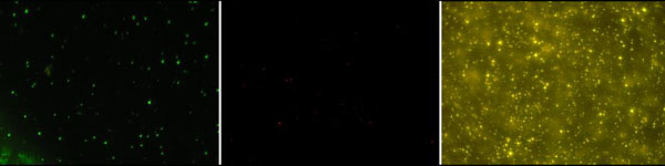
Fluorescent images of fluorophore-labeled liposomal vesicles.
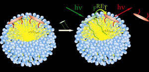
3. Using carefully selected dye molecules with extra-high FRET efficiency, the tags emission spectrum can be turned so that only the longest-wavelength dye will exhibit significant fluorescence at a short-wavelength excitation. This feature will overcome the challenge of small Stokes shift of many organic dyes. On the other hand, using dyes with different FRET efficiency, bar-code tags can be produced for multiplexed targeted FRET under single wavelength excitation.
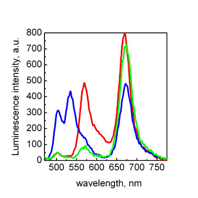
Fluorescence spectra of different dye compositions incorporated in nano-scale objects (liposomal vesicles) that demonstrates the bar-coding idea. Using specially selected dyes with high efficiency of FRET, fluorescent tags with an extremely large “Stokes shift” can be obtained.
4. Using FRET-tagging liposomes permits living cell – liposomes interaction to be visualized and studied in real time in vitro experiment. In this case, a loss of FRET signal could be used as a “signal system” to monitor liposome-cell fusion and active compound release.
Fluorescent images of rat hepatocytes incubated with FRET liposomes during different time periods and fluorescence spectra recorded in corresponding time period: 1 – FRET liposomes without cells, 2 – 0,5 h, 3 – 1h, 4 – 3h, 5 – 20h.
Main publications:
• A. Lebed, S. Yefimova, G. Guralchuk, A. Sorokin, I. Borovoy, Yu. Malyukin. Effect of hydrophobicity of cationic carbocyanine dyes DiOC n on their binding to anionic surfactant micelles//Journal of Applied Spectroscopy – 2010.– v.77., № 2. – P.183-188. abstract
•Yu.V. Malyukin, S.L. Yefimova, A.N. Lebedenko, A.V. Sorokin and I.A. Borovoy. Nano-scale control of energy transfer in the system “donor–acceptor”// Journal of Luminescence.– 2005.– v.112.– P.439-443. abstract
• Yu.V. Malyukin, S.L. Efimova, and K. Kemnitz. Spectroscopy of intermolecular interaction in the system: Dye–sodium dodecylsulphate micelles// Journal of Luminescence.– 2001.– v.94-95.– P.239-242. abstract
to the top